- +919347179399
- contact@thetoothstoryhyd.com
- Mon-Sat: 9:00am-9:00pm | Sun: 9:00am-1:00pm
Protect your oral and overall health with precise surgical removal of abnormal growths, supported by comprehensive pathological analysis.
Book Appointment
Cyst and tumor removal is a surgical procedure to excise abnormal growths that develop in the jaw, gums, oral soft tissues, or surrounding structures. These growths can be benign (non-cancerous) or, in rare cases, malignant (cancerous). The primary goals of treatment are to remove the abnormal tissue completely, preserve healthy structures, and obtain a definitive diagnosis through pathological examination.
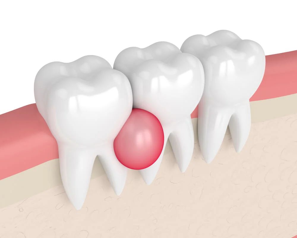
Cysts are fluid-filled sacs that can develop in the jawbone or soft tissues. While often benign, they can grow over time, destroying surrounding bone and damaging adjacent teeth.
Tumors are abnormal growths of tissue that may be benign or malignant. Early diagnosis and complete removal are critical for optimal outcomes.
All tissue removed during surgery is sent to a pathology laboratory for microscopic analysis, providing a definitive diagnosis and guiding any further treatment if needed.
Clinical examination, panoramic X-ray, CBCT (3D imaging), and possibly biopsy to actively assess the size, location, and nature of the growth.
Local anesthesia, IV sedation, or general anesthesia depending on the vast size and specific location of the growth.
Precision removal of the cyst or tumor, often securely including a clean margin of healthy tissue to verify complete removal.
Placement of advanced bone graft material if the cyst or tumor has unfortunately created a significant structural defect in the jawbone.
Submission of the completely removed tissue to an expert pathology laboratory for thorough, precise microscopic examination.
Discussion of final pathology remarkably results, ongoing monitoring for reliable recurrence, and careful evaluation of healing.
Early identification and completely precise removal of oral cysts and tumors are uniquely critical for your long-term oral and overall health.
The surgical experience varies based on the size, location, and nature of the growth.
Your oral surgeon will conduct a thorough examination, review imaging studies, and may perform a biopsy to obtain a preliminary diagnosis. The surgical plan is developed based on the size and location of the growth.
You will receive pre-operative instructions, including fasting guidelines if sedation or general anesthesia is planned. Any necessary pre-operative testing or medical consultations will be arranged.
Anesthesia is administered based on the complexity of the case. Local anesthesia may suffice for small superficial lesions, while larger or deeper lesions may require sedation or general anesthesia.
The surgeon makes an incision to access the growth, carefully removes it along with a margin of healthy tissue (if indicated), and preserves surrounding structures such as nerves, adjacent teeth, and vital anatomy.
If the removal creates a significant defect, bone grafting or other reconstructive techniques may be performed to restore form and function.
The surgical site is closed with sutures. You will rest in a recovery area before being discharged with detailed aftercare instructions. Once the pathology report is available (typically 5–10 days after surgery), your surgeon will discuss the results with you and monitor for signs of recurrence.
Surgical removal is indicated when a cyst or tumor is identified in the oral or maxillofacial region.